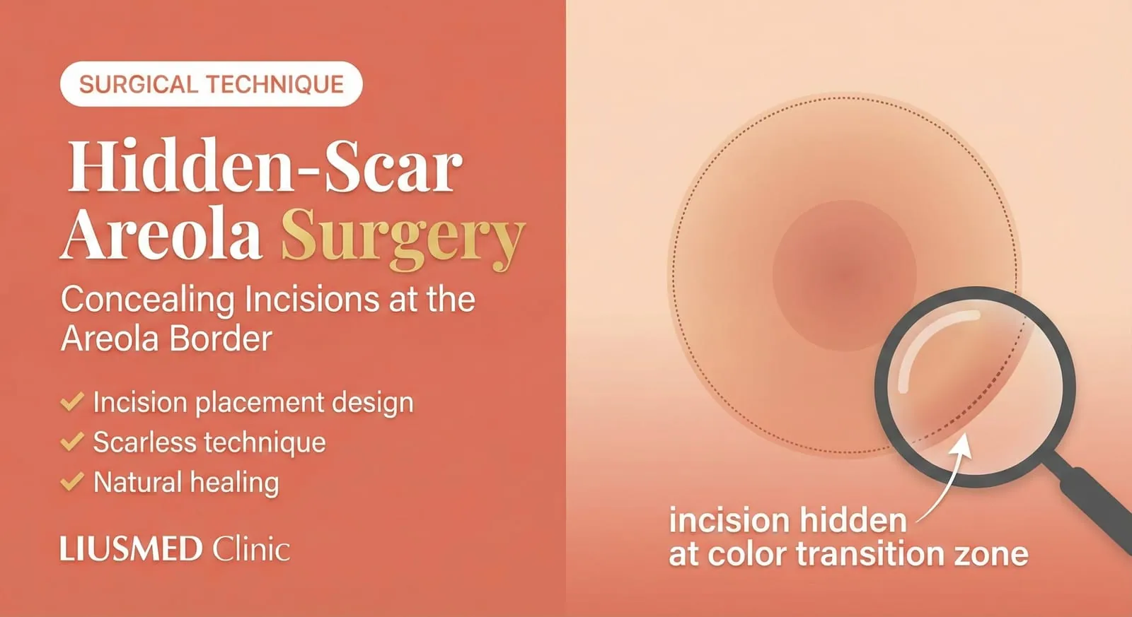
The Core Concept of Hidden-Scar Surgery
The biggest challenge in areola odor surgery is how to eliminate odor without leaving visible scars. The answer lies in precise incision design—placing surgical incisions along the "color boundary line" between the areola and normal skin. After healing, the scar is concealed by the natural color difference, achieving near-invisible results.
Anatomical Features of the Areola
Why Is the Areola Ideal for Hiding Scars?
| Feature | Explanation |
|---|
| Clear color contrast | Areola is darker, with distinct boundary from surrounding skin |
| Natural texture | Fine surface texture helps conceal small scars |
| Thinner skin | Healed incisions have fine lines |
| Good blood circulation | Fast healing, low scar formation risk |
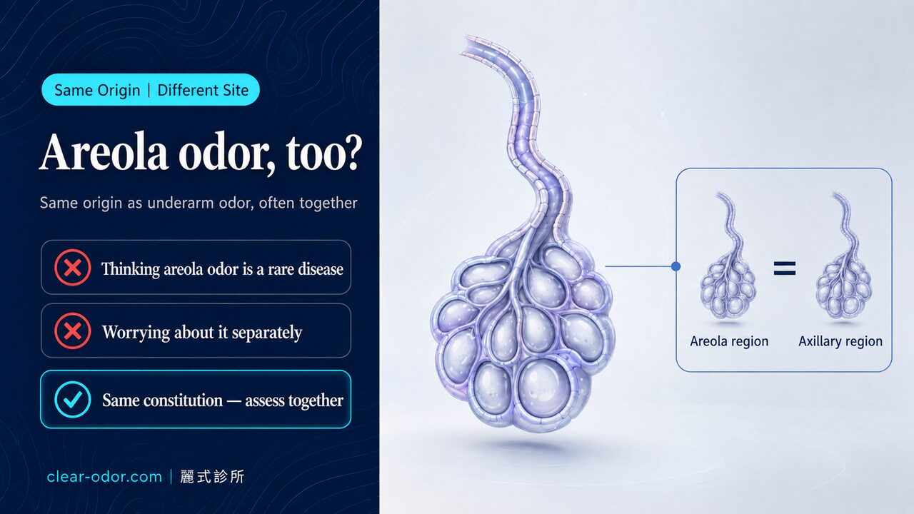
Distribution of Odor Glands in the Areola Area
Areola odor apocrine glands are mainly distributed in:
- Outer periphery of areola: Highest concentration
- Between areola and nipple: Medium density
- Around nipple: Lower density
💡 Dr. Liu's Observation: "The distribution area of areola apocrine glands is usually much smaller than the underarm, meaning the surgical area is relatively limited—making thorough removal without leaving marks even more achievable."
Three Principles of Incision Design
Principle 1: Design Along the Color Boundary
The golden rule of incision design is overlapping the incision line with the areola edge:
| Incision Position | Post-Op Scar Visibility |
|---|
| Upper areola (12 o'clock) | Low (covered by bra) |
| Lateral areola (3 or 9 o'clock) | Very low (hidden on side) |
| Lower areola (6 o'clock) | Very low (hidden below) |
| Areola edge color boundary | Nearly invisible |
Principle 2: Follow Skin Tension Lines
Skin has natural tension lines (Langer's lines). Cutting along these lines:
- Minimizes tension during healing
- Creates finest, flattest scars
- Reduces hypertrophic scarring risk
Principle 3: Minimize Incision Length
The essence of minimally invasive technique is completing the maximum treatment area through the smallest incision:
| Surgical Method | Incision Length | Treatable Area |
|---|
| Traditional Excision | 3-5 cm | Fixed range |
| Minimally Invasive Curettage | 0.5-1 cm | Extended treatment range |
Technical Details of Hidden-Scar Surgery
Step 1: Pre-Operative Marking
- Mark apocrine gland distribution: Use iodine testing or palpation to identify odor source area
- Design incision position: Choose the most concealed area at areola edge
- Confirm symmetry: Left and right incision positions should be symmetric
Step 2: Creating the Incision
| Step | Technical Points |
|---|
| Local anesthesia | Tumescent technique reduces bleeding |
| Incision | Along areola edge color line, blade perpendicular to skin |
| Depth | Only through epidermis and dermis, not into deep layers |
| Length | 5-10 mm is sufficient |
Step 3: Apocrine Gland Curettage
Through the small incision, specialized instruments are used to curette the apocrine gland layer:
Incision → Create subcutaneous tunnel → 180-degree fan-shaped curettage → Confirm complete removal
Technical Keys:
- Maintain correct anatomical layer (subdermal)
- Avoid going too deep into fat layer or mammary tissue
- Avoid going too superficial, causing skin necrosis
Step 4: Closure and Suturing
| Suturing Technique | Effect |
|---|
| Subcutaneous sutures | Reduces skin tension |
| Fine skin sutures | Using 6-0 or 7-0 fine thread |
| Precise alignment | Ensures smooth areola edge |
| Eversion sutures | Reduces depressed scarring |
Why Does the Scar Become "Invisible"?
Visual Principles Explained
Scar invisibility is based on human visual characteristics:
| Factor | Explanation |
|---|
| Color contrast concealment | Color difference between areola and skin distracts attention |
| Texture blending | Small scar blends into natural areola texture |
| Hidden location | Areola edge is not a visual focal point |
| Normal coverage | Usually covered by undergarments |
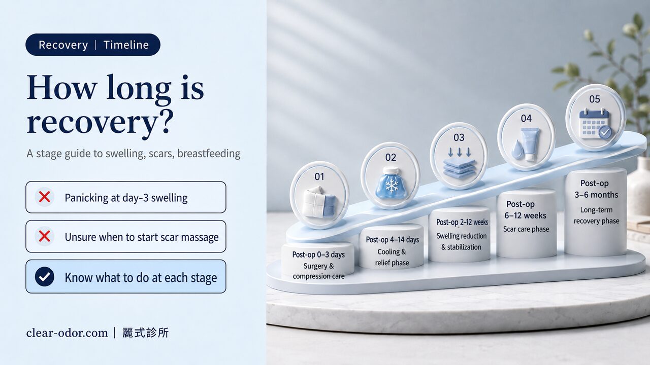
Post-Op Scar Evolution Timeline
| Period | Scar Status |
|---|
| Week 1 | Suture site slightly red, clearly visible |
| Month 1 | Scar begins fading, fine line still visible |
| Month 3 | Scar significantly faded, blends with areola edge |
| Month 6 | Nearly invisible, must look carefully to notice |
| Year 1 | Completely blended, cannot identify incision location |
💡 Dr. Liu's Experience: "By about 3-6 months post-op, most patients have difficulty finding their original incision location themselves. This is the advantage of the color boundary incision."
Incision Strategies for Different Areola Types
Adjusting for Areola Size
| Areola Type | Incision Strategy |
|---|
| Small areola (< 3cm) | Single incision, enter from lateral side |
| Medium areola (3-4cm) | Can choose single or bilateral incisions |
| Large areola (> 4cm) | May need two symmetrical incisions |
Adjusting for Areola Color
| Areola Color | Incision Design |
|---|
| Dark areola | Clear color contrast, best scar hiding effect |
| Light areola | Less contrast, requires finer suturing technique |
| Irregular border | Design along natural edge, hiding with contour |
Comparison with Other Techniques
Incision Location Comparison
| Surgical Method | Incision Location | Scar Visibility |
|---|
| Traditional direct excision | Center of areola | Obvious |
| Upper areola incision | Upper edge of areola | Medium |
| Lateral areola incision | Lateral areola | Low |
| Color boundary incision | Areola edge | Minimal |
Post-Op Results Comparison
| Item | Traditional Surgery | Hidden-Scar Minimally Invasive |
|---|
| Incision length | 3-5 cm | < 1 cm |
| Scar width | 3-5 mm | < 1 mm |
| Recovery time | 2-3 weeks | 1 week |
| Scar maturation | 6-12 months | 3-6 months |
Post-Op Scar Care
Methods to Accelerate Scar Invisibility
Phase 1 (Weeks 1-4 Post-Op)
| Care Item | Method |
|---|
| Cosmetic tape | Apply along wound direction, change every 3-5 days |
| Sun protection | Avoid direct sunlight |
| Cleanliness | Keep dry, prevent infection |
| Care Item | Method |
|---|
| Silicone sheets | Use at least 12 hours daily |
| Scar massage | After wound fully healed, gentle daily massage |
| Continue sun protection | Prevent pigmentation |
| Care Item | Method |
|---|
| Silicone gel | Can switch to topical products |
| Observation | Address any abnormal growth early |
Extra Care for Special Constitutions
| Constitution | Recommendations |
|---|
| Keloid tendency | Extend silicone use to 6-12 months |
| Darker skin tone | Enhanced sun protection, prevent pigmentation |
| Dry skin | Use moisturizing products to maintain skin elasticity |
Frequently Asked Questions
Q1: Will the incision be on the areola?
A1: No. The incision is designed at the boundary between areola and normal skin, not on the pigmented areola area. This way, the healed scar is concealed by the color difference.
Q2: Will both sides have incisions in the same location?
A2: Symmetrical positions are designed, usually at the lateral or lower edge of the areola color boundary. This ensures both sides look symmetrical and natural.
Q3: If my areola color is very light, will the scar be more visible?
A3: Light-colored areolas do have less color contrast effect than dark areolas, but with correct incision placement and fine suturing, scars still become very inconspicuous by 3-6 months.
Q4: Can I sunbathe after surgery?
A4: Avoid direct sunlight on the scar for 3 months post-op—UV rays cause pigmentation, making scars darker. Though the areola area is usually covered by undergarments, be mindful when wearing swimwear.
Q5: If I'm not satisfied with the scar, are there remedies?
A5: After the scar stabilizes at 6 months, if there's still a slight trace, options include:
- Laser treatment: Improves color difference and texture
- Microneedling: Stimulates collagen remodeling
- Injection treatment: For hypertrophic scars
Most cases don't require additional treatment.
Surgical Process Overview
- Pre-Op Evaluation → Design Incision Location → Local Anesthesia
- Cut Along Color Line → Create Subcutaneous Tunnel → Curette Apocrine Glands
- Confirm Complete Removal → Fine Suturing → Post-Op Care Instructions
- Suture Removal at 1 Week → Begin Scar Care → Regular Follow-Up
Advantages of Choosing Hidden-Scar Technique
| Advantage | Explanation |
|---|
| Aesthetic | Scar nearly invisible |
| Confidence | No worry about visible scars |
| Quick recovery | Small wound, fast healing |
| Safe | Minimally invasive, low risk |
| Effective | High odor elimination rate |
Related Reading
Clear Odor Specialist Perspective
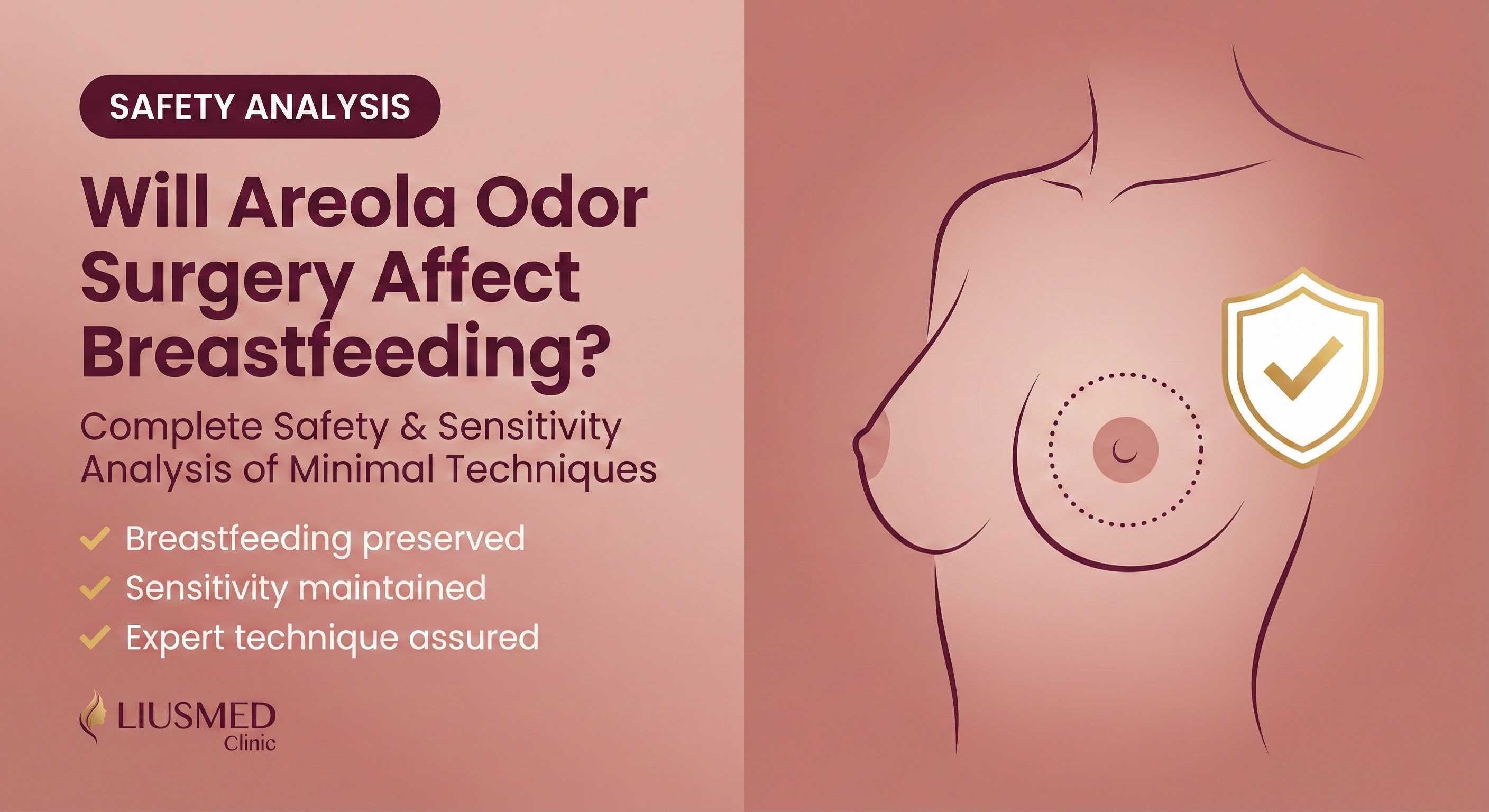
Clear Odor Clinic is dedicated exclusively to body-odor and hyperhidrosis treatment, and areolar bromhidrosis is one of the more technically demanding categories we see. Every patient receives an individual assessment of areolar anatomy, breastfeeding history, and family-planning context — and Dr. Ta-Ju Liu personally plans the incision rather than applying a standardised axillary protocol.
Years of dedicated focus on areolar cases have let us refine a tighter safety margin: the mammary and ductal systems are strictly preserved, only the superficial-dermis apocrine layer is addressed, and post-operative high-tension movement is restricted for 6 months to protect the areolar scar. These are details only an odor-focused clinic has the case volume to polish.
Full Treatment Information → · Book a Specialist Consultation →
Related Reading
About the Author
Dr. Ta-Ju Liu
- Current Position: Director, Liushi Clinic
- Specialties: Minimal incision surgery (lipoma, cyst), hyperhidrosis surgery, thread lifting
- Experience:
- 15+ years of clinical minimal incision surgery experience
- Over 10,000 successful minimal incision cases
- Board-certified dermatologist
- Philosophy: "Scar invisibility is an art. Every incision position and angle is carefully designed. My goal is for patients to completely forget they ever had surgery."